Extreme macro: a freshly cut kiwi slice, about 2 mm in thickness, under the microscope
The kiwi (actinidia deliciosa) is a fascinating fruit with a complex anatomy and intriguing structures, making it a popular subject for photography.
We captured minute details of this alluring fruit using two of our devices – the LM macroscope and the LM photomicroscope – and high-resolution microscope lenses. These devices, which are optimised for photomicrography and photomacrography applications, make it possible to use standard microscope lenses and special lenses with mirrorless system cameras and DSLRs.
Microscope lenses are among the most powerful optical lenses for capturing images of the smallest microscopic structures, as they are designed for maximum resolution and light gathering power. The numerical aperture (NA) is the key metric that determines the system’s ability to gather light and resolve details.
To achieve greater depth of field, we used focus stacking: we took 10-20 individual images, combined them into a stack and processed them with the Helicon Focus image processing software. We created our stack using a motorised stage and the popular StackShot focusing rail, moving the camera/lens unit along the z-axis. To create a transmitted illumination source, we used an LED light box, illuminating the entire specimen and revealing the intricate structures within the 2 mm thick section.
For our photography setup, we used two different cameras: an older Nikon D610 DSLR (manufactured in 2014) with a full-frame sensor, and a Canon R5 mirrorless system camera (built in 2023). The Nikon D610, even though it is more than 10 years old, impresses with its 24-megapixel sensor’s ability to produce high-quality images with a wide dynamic range.
Exquisite, green, aesthetic images:
The lush, vivid green beautifully offsets the seeds and the white-green, fibrous pulp of the fruit, interwoven with strands and fibres. The kiwi’s thin and brown outer skin is often covered with fine, hair-like textures. It protects the fleshy interior of the fruit and stands in stark contrast to the vibrant green pulp speckled with black seeds.
Image: kiwi slice, overview shot with LM macroscope and Nikon D610, StackShot, Helicon software, stack of 20 individual images:
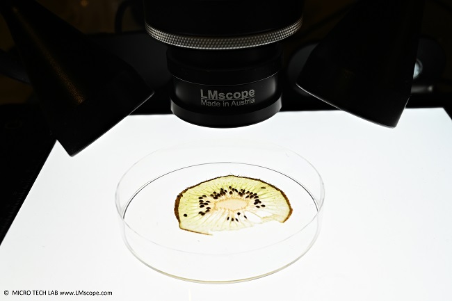
We cut the specimen both lengthwise and crosswise, producing thin slices approximately 2 mm in thickness. Attempting to get any thinner cuts with a standard kitchen knife doesn’t really work, as the slices tend to disintegrate, especially considering the 1-2 mm size of the seeds. For storage and placement, we put the kiwi slice in a Petri dish.
Image: overview of thin kiwi slice in a Petri dish with white LED backlighting
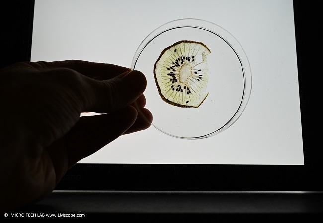
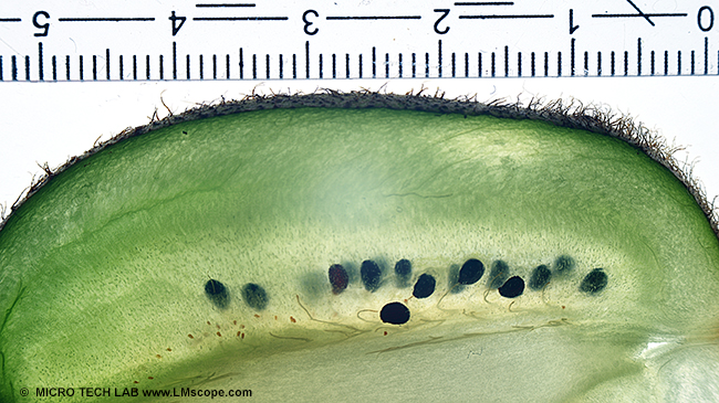
Image below: overview shot of a kiwi slice cut lengthwise. For this image, the specimen was placed directly onto the light box, next to a standard ruler with millimetre markings. The Kaiser LED light box is spacious enough to accommodate even large specimens.
If documentation is required, placing a standard ruler at the edge of the image allows for future calibration of the measurement software, ensuring precise measurement of the image details.
For this shot, we used an older full-frame Nikon D610 camera in combination with the LM macroscope, equipped with a Nikon F-mount bayonet lens mount in this instance.
Example application: LM photomicroscope, LED light box and two LED spots
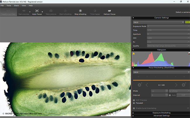
In this example application, we equipped the stage with a motorised StackShot focusing rail. To automate the process of taking a sequence of images, we used the powerful Helicon Remote software, which controls both the camera and the motorised stage. This allows the camera/lens unit to be moved along the z-axis via computer control, automating the capture of a larger stack of images taken at different focus distances.
Example application: LM macroscope stage with StackShot rail, attached directly to the camera body. This mounting method enables quick lens changes for fine-tuning the magnification or field size.

We used a standard USB cable to connect both camera and stage to the StackShot control unit. The Helicon software displays the Live View window on the computer and gives full control over all settings of the camera and stage. The two focusing endpoints can also be set from computer – a convenient feature.
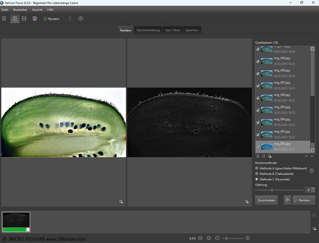
After the stack has been created, the next step is to start the Helicon Focus processing software to process the individual images and merge them into one composite image.
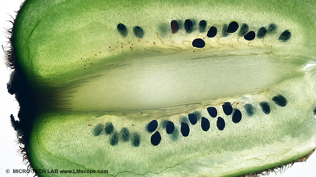
A closer look at the seeds and pulp:
Next, we used the LM wide-field photomicroscope module to display the intricate structures of the kiwi at a higher magnification. This time, an LED transmitted light unit served as illumination source.
The tiny seeds are embedded in the fruit’s pale green, milky-translucent pulp. The pulp itself is reinforced by small fibrous structures. Delicate threads extend to the base of the seeds, supplying them with essential nutrients during the kiwi’s growth phase.
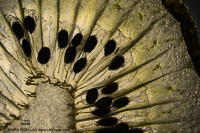
Image: kiwi slice, close-up at 30x microscope magnification with LM wide-field photomicroscope, StackShot, Helicon software, stack of 20 individual images
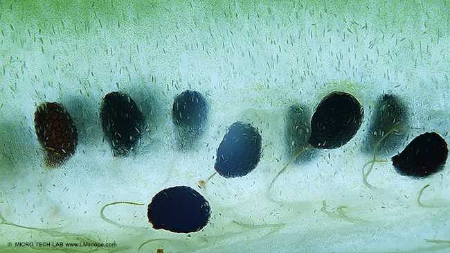
Image below: close-up of the seed structures in polarised light (transmitted light). The fibrous structures within the pulp are distinctly visible. The surface of the seeds with its three-dimensional structure appears very dark.
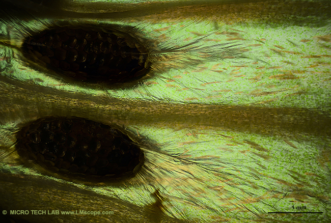
Image below: close-up of the kiwi slice. LED illumination with a combination of transmitted and side lighting, LM wide-field photomicroscope, 100x microscope magnification, Canon EOS R5
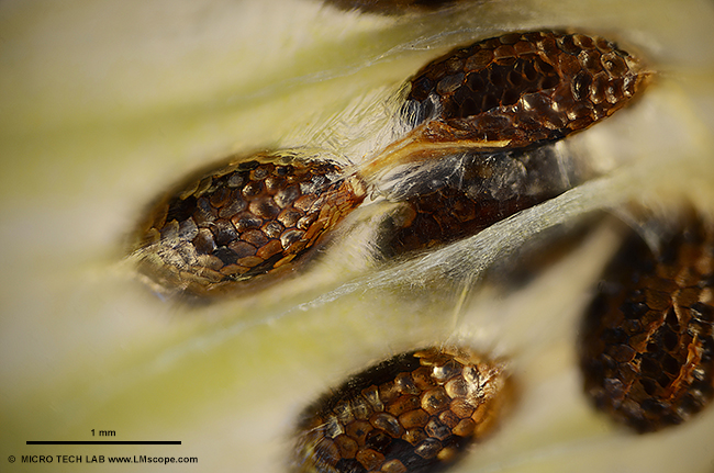
Image below: close-up of kiwi cross-section, LED transmitted light, polarisation
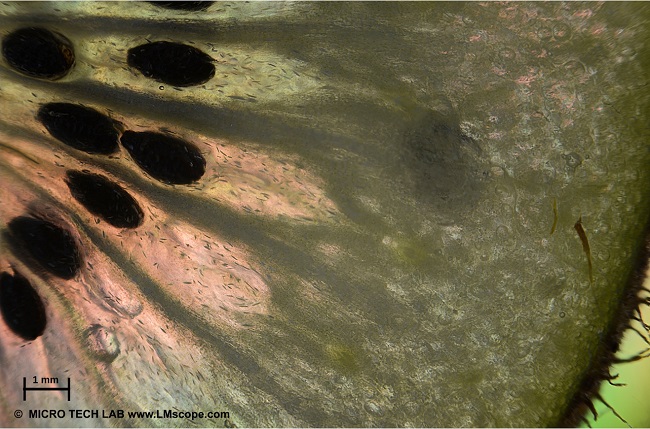
Image below: close-up of kiwi cross-section, LED transmitted light / darkfield